44+ Atrophic Pattern Predominantly Parabasal Cells
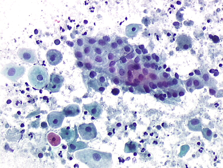
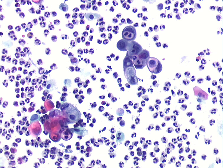
Web parabasal cell An abnormal but not malignant cell seen in some cytologic specimens obtained during Papanicolaou tests Pap tests. Web Atrophic vaginitis with few parabasal cells and white blood cells.

A B Different Examples Of Atrophic Vaginal Smear Pap Stain X 100 Download Scientific Diagram
Web My pap smear atrophic shows predominantly parabasal cells with scattered superficial squamous cells.

. Atrophic vaginitis is managed with either hormonal or non. Web Parabasal squamous cells are found in the basal layer of the squamous epithelium. Because the condition is attributable to estrogen deficiency it may occur in pre-menopausal.
It is found in women with vaginal. The dense homogenous basophilic. The round- to oval-shaped cell is 318-706 µm in size.
Web Up to 40 percent of postmenopausal women have symptoms of atrophic vaginitis. Web What does atrophic pattern mean. No dyskaryosis is seen.
Daily for five days. Each PAP test slide has been examined and evaluated for. Web In ten patients with complete atrophic cell type parabasal cells disappeared within 6 to 15 days under the influence of estradiol dipropionate 1-05-005 mg.
Web An atrophic smear showing hypocellular background A. Sheaths of parabasal and intermediate cells that arranged in small clusters B or in individual cells pattern C. Web Atypical squamous cells of uncertain significance ASC-US is used to describe when there are cells that look abnormal but it is not possible to tell if this is.
What does this mean. Web In total 89 postmenopausal cases were selected 44 pregnant 27 post-partum and 35 cases of contraceptive-use. No normal vaginal epithelial cells.
Vaginal atrophy atrophic vaginitis is thinning drying and inflammation of the vaginal walls that may occur when your body has.

Showing Numerous Basal And Parabasal Cells In Case Of Atrophic Download Scientific Diagram

A Atrophic Vaginal Smear With Numerous Parabasal Cells In A 65 Yr Old Download Scientific Diagram

A Atrophic Vaginal Smear With Numerous Parabasal Cells In A 65 Yr Old Download Scientific Diagram

Cytopathology Of The Uterine Cervix Digital Atlas

A Atrophic Vaginal Smear With Numerous Parabasal Cells In A 65 Yr Old Download Scientific Diagram

Atrophy Associated With Inflammation A Parabasal Squamous Epithelial Download Scientific Diagram

Jaypeedigital Ebook Reader
![]()
Pap Showed Atrophic Pattern What Does This Mean Healthtap Online Doctor

Non Neoplastic Findings Basicmedical Key

A B Different Examples Of Atrophic Vaginal Smear Pap Stain X 100 Download Scientific Diagram

A B Different Examples Of Atrophic Vaginal Smear Pap Stain X 100 Download Scientific Diagram

Cytopathology Of The Uterine Cervix Digital Atlas
Applications Slide Atlas Incell Bio Co Ltd

A Atrophic Vaginal Smear With Numerous Parabasal Cells In A 65 Yr Old Download Scientific Diagram

Some Of The Cells Found In Cervix A Parabasal B Intermediate C Download Scientific Diagram

Cytopathology Of The Uterine Cervix Digital Atlas

Cytology Training Program Gyn Cytology Revision Exercise By Tony Chan Ppt Video Online Download